
The uniformity in light output of pure CsI crystals was measured in a test setup with a 137Cs radioactive source. It was found that this method is consistent with the 3D tomography and well suited for uniformity tests. Thus an apparatus has been designed which allows a fast, precise and reproducible method to determine the optical non-uniformity of each CsI crystal.
The 137Cs-source is embedded in a Pb-collimator. The collimator has a thickness of 5 cm; the probability for the 0.66 MeV photons to penetrate the lead is <1%. Its opening has a diameter of 6 mm. The collimator is mounted on a plate, which is moved by an ISEL stepping motor. The precision of the step motor is ~12.5 microns. The crystal is placed on an aluminum plate using a dedicated positioning system with a precision of ~0.3mm. The step motor is PC controlled using the RS232 interface.
The signals from the PMT are amplified by a factor of 10 using a LRS 612A PM-Amplifier. The signal is then split, one part going in the trigger the other is delayed and fed into a LRS 2249A CAMAC ADC. The trigger creates a computer LAM in the IO506 unit. The CAMAC modules are controlled using a HYTEC 1331 Turbo CAMAC-PC interface.
The main program to operate the apparatus is called RASTA. Its main tasks are:
On execution of the program, it asks first for the serial number sxxx of the crystal to be tomographed. A new directory is created \hisxxx in which finally all the relevant histograms of the scan process are stored.
The starting parameters of the program are defined in the file
RASTA.INI:
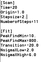
The category Scan controls the scanning process:
The category Fit controls the fitting process:
After having changed or accepted the run parameters the step motor is initialized and the collimator is moving to the parking position where pedestal data and background data are taken.
Then, the crystal is scanned at the points specified in RASTA.INI. The following picture shows the display as it appears during the process of scanning.
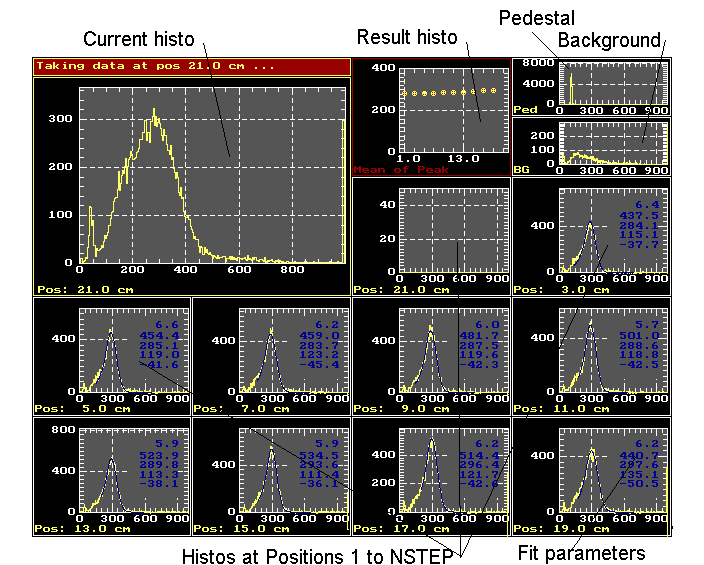
As results the data of all the histograms are stored and can be easily viewed with EXCEL (Result histos 0 and 1) and Histo-manager (Histos 2 to NSTEP+3).
Two other programs are supplied :
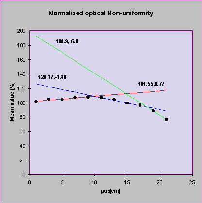
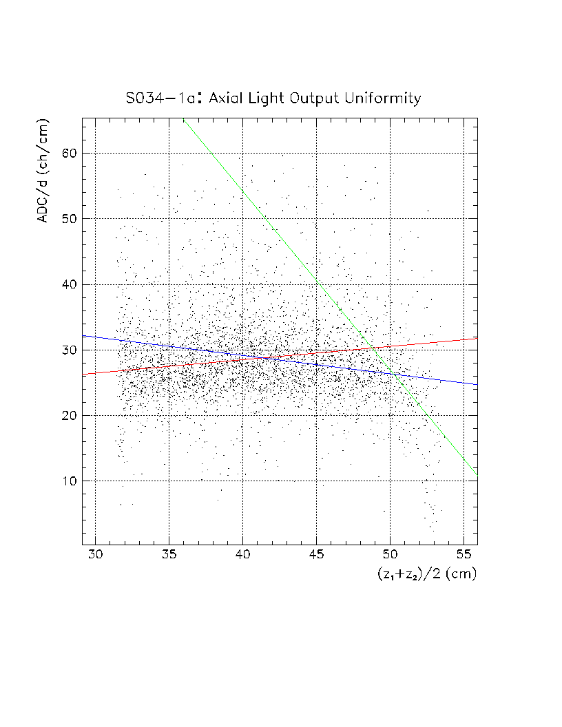
The resulting optical non-uniformity obtained with RASTA can be compared very easily to the cosmic ray tomography results:
The resulting parameters for s034 are:
| Method | Slope 0:10 cm | Slope 10:18 cm | Slope 18:22 cm |
| RASTA | 0.77 %/cm | -1.88 %/cm | -5.8 %/cm |
| CRT | 0.69 %/cm | -0.96 %/cm | -11.261 %/cm |
The resulting slopes, especially for the first 10 cm, agree very well for the two methods.
The stability of the system after switching on the high voltage has been determined. Data were taken at the same position every 5 minutes during one hour:

As a conclusion, the high voltage of the crystal should be switched on for at least 40 minutes before doing any measurements with the crystal.
A full scan has been performed on a crystal 4 times within 3 hours. The obtained results for s040 are shown in the figure below:
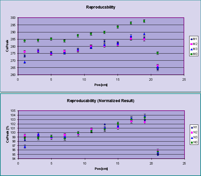
After normalization the results of different measurements agree
within the errors.